Patent Ductus Arteriosus (PDA) in Dogs and Cats
What is patent ductus arteriosus?
Patent ductus arteriosus (PDA) is one of the most common congenital heart defects in dogs. Together with subaortic stenosis and pulmonic stenosis, it belongs to the most frequently diagnosed congenital cardiac diseases in veterinary cardiology.
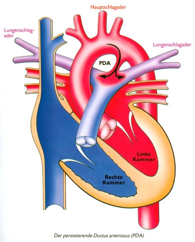
Before birth, the ductus arteriosus is a normal fetal blood vessel connection between the aorta and the pulmonary artery. After birth, this vessel should close spontaneously. If it remains open, it is called a patent ductus arteriosus.
As a result, blood flows abnormally from the aorta back into the pulmonary circulation, which can overload the heart and eventually lead to heart failure if left untreated.
Which dogs and cats can be affected?
PDA can occur in many breeds and can also be diagnosed in cats. Breeds reported more commonly include:
| Miniature and Toy Poodle | Collie | Pomeranian |
| German Shepherd Dog | Cocker Spaniel | Shetland Sheepdog |
| Old English Sheepdog | Maltese | Yorkshire Terrier |
| Rottweiler | Keeshond |
What happens in a patient with PDA?
After birth, pressure in the aorta is normally higher than in the pulmonary artery. Therefore, in a left-to-right PDA, oxygenated blood flows from the aorta back through the open ductus into the pulmonary artery.
This causes pulmonary overcirculation and volume overload of the heart, especially of the left atrium and left ventricle.
Why is PDA important?
Untreated PDA can cause serious long-term damage and in most cases should be closed as early as possible.
Possible consequences: heart enlargement, heart failure, pulmonary oedema, pulmonary hypertension
Important: if a right-to-left shunt develops, closure is generally contraindicated
Some dogs may appear clinically stable at first, but the abnormal blood flow can still damage the heart and lungs over time.
What symptoms can occur?
Many dogs with a left-to-right PDA initially show few or no obvious symptoms. Without treatment, however, many patients will eventually develop congestive heart failure.
Typical signs of left-sided heart failure include:
- Fast or difficult breathing
- Coughing
- Exercise intolerance
If a right-to-left shunt develops, signs may include:
- Bluish mucous membranes, sometimes as differential cyanosis
- Weakness or collapse of the hind limbs during exercise
- Fainting episodes
- Lethargy
How is PDA diagnosed?
The clinical examination often provides very important clues. An experienced veterinarian or cardiologist may strongly suspect PDA based on the typical continuous heart murmur alone.
A very strong peripheral pulse is also common. In some patients a palpable precordial thrill may be present.
What does a PDA murmur sound like?
PDA typically causes a continuous murmur, often described as a “machinery murmur”, heard best over the left heart base.
You can also listen to the audio file directly here: PDA murmur audio sample.
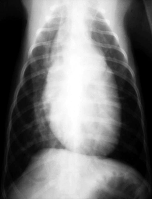
What do chest radiographs show?
Radiographic findings depend on the size of the ductus. Small PDA may cause only mild or no radiographic changes. Larger defects often result in cardiomegaly and signs of increased pulmonary blood flow.

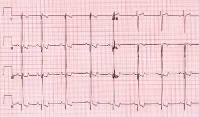
What can the ECG show?
Typical ECG findings may include large R waves caused by left ventricular enlargement and volume overload.
What does echocardiography show?
Echocardiography is the key diagnostic tool for PDA. It usually shows volume overload of the left atrium and left ventricle.
Doppler imaging is used to assess blood flow through the ductus and to estimate the pressure gradient between the aorta and the pulmonary artery.
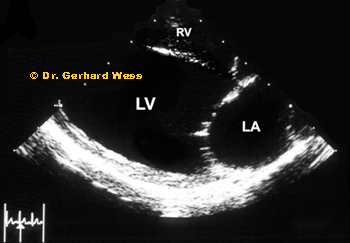 Typical findings include dilation of the left ventricle and enlargement of the left atrium.
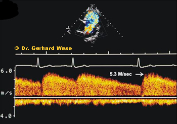 Continuous-wave Doppler helps estimate the velocity through the PDA and assess whether pulmonary hypertension may already
be developing.
Continuous-wave Doppler helps estimate the velocity through the PDA and assess whether pulmonary hypertension may already
be developing.
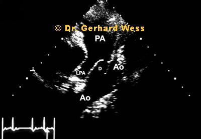 In many patients, the ductus itself can also be directly visualised, which helps assess its anatomy and plan treatment.
To better understand the blood flow pattern, here is a colour Doppler example:
What is the prognosis?
If PDA is closed early and successfully, prognosis is usually excellent.
Without treatment, many patients eventually develop heart failure and may die at a young age. After successful closure, many dogs can have a normal life expectancy.
How is PDA treated?
In most patients, PDA should be closed, ideally before irreversible heart or lung damage has developed. Today, this is often done by a minimally invasive catheter-based procedure.
The catheter is usually advanced through a femoral artery to the heart. This is much less invasive than open thoracic surgery and is generally the preferred approach when anatomically possible.
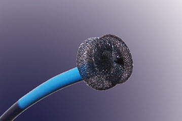 A commonly used closure device is the Amplatzer Duct Occluder, which is positioned within the ductus by catheter.
 In smaller patients or in selected anatomical situations, a coil-based procedure may also be used.
Surgical ligation remains an option, especially in very small patients or when the ductus anatomy is not suitable for catheter closure.
PDA closure at LMU Munich
We have a separate page dedicated specifically to the interventional PDA closure procedure.
This page explains the catheter-based treatment, device selection and the differences compared with surgical closure.