Heart Murmurs in Dogs and Cats
Important information for pet owners
Heart murmurs are often the first clue that a dog or cat may have heart disease. They are commonly detected during a routine examination, vaccination visit or breeding check.
It is important to know that not every murmur means severe heart disease. On the other hand, not every relevant heart disease produces an obvious murmur.
Because the underlying cause usually can only be defined reliably with echocardiography, a newly detected murmur should be assessed carefully.
What should owners know?
A murmur is not a final diagnosis. It is an abnormal sound produced by altered blood flow in the heart or great vessels.
These turbulent flow sounds may be harmless, but they can also occur with congenital or acquired heart disease. This is why careful cardiac work-up is especially important in puppies, kittens, breeding animals, and older pets with a newly detected murmur.
Important: Not every murmur is dangerous.
Also important: Without echocardiography, a relevant heart problem often cannot be ruled out reliably.
What is auscultation?
Auscultation means listening to the heart and lungs with a stethoscope. It is a central part of every cardiovascular examination.
An experienced examiner can obtain important clues about heart rate, rhythm, additional heart sounds and murmurs. Some heart diseases produce such typical murmurs that a strong suspicion can already arise from auscultation.
During auscultation, the pulse should also be checked. If not every heartbeat produces a palpable pulse wave, this is called a pulse deficit and may indicate an arrhythmia.
Important auscultation findings
- abnormal heart rate, such as bradycardia or tachycardia
- irregular rhythm
- abnormal intensity of heart sounds
- additional heart sounds
- split heart sounds
- heart murmurs
Heart sounds and murmurs – what is the difference?
Normal heart sounds are produced mainly by valve closure. A heart murmur is an additional abnormal sound caused by turbulent blood flow.
In small animals, two main heart sounds are usually heard:
- First heart sound (S1): closure of the mitral and tricuspid valves
- Second heart sound (S2): closure of the aortic and pulmonic valves
Under certain circumstances, a third heart sound (S3) or fourth heart sound (S4) may also be heard. If extra sounds are present in addition to S1 and S2, this is called a gallop rhythm.
Muffled heart sounds may occur with obesity, pleural effusion, pericardial effusion or thoracic masses. Clearly changing heart sound intensity often points toward arrhythmias.
What types of murmurs are there?
Heart murmurs are classified as systolic, diastolic or continuous.
The vast majority of murmurs in small animal cardiology are systolic murmurs.
Systolic murmurs
Systolic murmurs are heard after the first heart sound and end before the second heart sound. They commonly occur with:
- mitral or tricuspid regurgitation
- aortic stenosis or subaortic stenosis
- pulmonic stenosis
- ventricular septal defect (VSD)
- other congenital defects such as tetralogy of Fallot
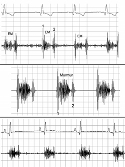
Diastolic murmurs
Diastolic murmurs are less common. They begin after the second heart sound. Typical causes may include:
- aortic valve insufficiency
- rare mitral or tricuspid stenosis
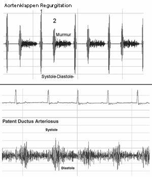
Continuous murmurs
A continuous murmur does not stop at the second heart sound. It is present in both systole and diastole. This is especially typical for a patent ductus arteriosus (PDA).
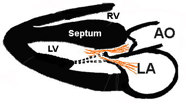
Where is a murmur heard most loudly?
The location of the point of maximal intensity often gives very important clues to the cause.
The most important area is where the murmur is heard most loudly on the chest wall. Together with the patient's age, breed and murmur type, this often helps narrow the list of possible diagnoses.
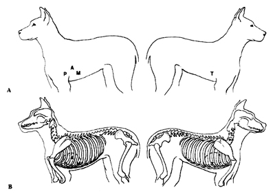
Location of the main valves
P = pulmonic valve, A = aortic valve, M = mitral valve, T = tricuspid valve
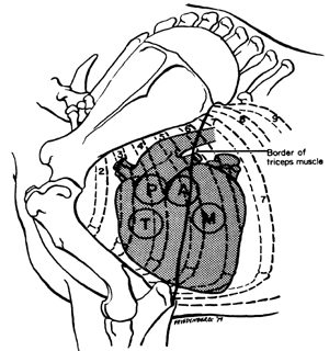
- Pulmonic and aortic valve: left cranial thorax, usually around the 3rd–4th intercostal space
- Mitral valve: left apical area, further caudal, usually around the 5th–7th intercostal space
- Tricuspid valve: right side, usually around the 4th intercostal space
The PABST rule
A simple memory aid is the PABST rule:
Pulmonic – Aortic – Bicuspid (= mitral) – Side change – Tricuspid
Common murmurs in dogs
- Aortic stenosis / subaortic stenosis: systolic murmur left heart base; in severe cases the pulse may be weak
- Pulmonic stenosis: systolic murmur left heart base
- Patent ductus arteriosus (PDA): continuous murmur left base, often with bounding pulses
- Mitral regurgitation: systolic murmur left apex
- Tricuspid regurgitation: systolic murmur right side
- Ventricular septal defect (VSD): usually systolic, often loudest on the right cranial chest
- Atrial septal defect (ASD): often no typical murmur; sometimes a systolic murmur is heard over the pulmonic area
How are murmurs graded?
Murmur intensity is commonly graded on a 1 to 6 scale:
- Grade 1: very soft, only heard after careful listening in a quiet room
- Grade 2: soft, but heard immediately
- Grade 3: moderate intensity
- Grade 4: loud, without a palpable thrill
- Grade 5: very loud, with a palpable thrill
- Grade 6: extremely loud, in some cases audible with only minimal contact
The grade alone does not determine the exact disease.
Special considerations in cats
In cats, the cause of a murmur is often more difficult to define from auscultation alone than in dogs.
Many feline murmurs are best heard parasternal, close to the sternum. Even for experienced examiners it is often difficult to assign the murmur precisely to one valve area.
For that reason, any cat with a murmur should ideally have an echocardiogram.
SAM in cats
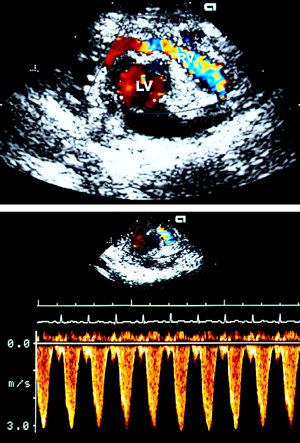
SAM stands for systolic anterior motion of the mitral valve. This murmur is seen especially in cats with hypertrophic cardiomyopathy (HCM).
Because of altered left ventricular geometry, part of the mitral valve moves toward the outflow tract during systole. This can cause a dynamic left ventricular outflow tract obstruction and often also mitral regurgitation. The murmur may be more obvious at higher heart rates and may disappear when heart rate slows.
Dynamic right ventricular outflow tract obstruction in cats
Dynamic right ventricular outflow tract obstruction, often referred to as dynamic RV outflow murmur, is another common feline murmur pattern.
In many cats, this is a rather benign dynamic murmur that is heard mainly at higher heart rates. In some cases it may also occur together with cardiac or systemic disease.
When should a murmur be evaluated further?
When in doubt, earlier evaluation is better.
- any newly detected murmur in a puppy or kitten
- murmurs detected during breeding examinations
- older dogs or cats with a newly developed murmur
- animals with cough, dyspnea, exercise intolerance, collapse or increased resting respiratory rate
- cats in general, because auscultation alone often cannot define the cause reliably
Which tests are useful?
The most important examination is echocardiography with Doppler. Depending on the case, the following may also be useful:
- Echocardiography
- ECG
- Holter monitor / 24-hour ECG
- Thoracic radiographs
Audio examples of heart murmurs
These examples are easiest to appreciate with headphones.
| Audio | Example |
|---|---|
 |
Normal auscultation: first and second heart sound, small dog over the apex |
|
Normal auscultation: first and second heart sound, small dog over the heart base |
|
Mild mitral regurgitation with a musical murmur |
|
Severe mitral regurgitation |
|
Chaotic rhythm with murmur: atrial fibrillation and mitral insufficiency |
|
Cat: normal auscultation |
|
Cat: gallop rhythm, S3 with HCM |
|
Cat: another example of gallop rhythm in HCM |
|
Cat: systolic murmur in hypertrophic cardiomyopathy with SAM |
|
Continuous murmur with PDA |
|
Another example of PDA |
|
Systolic murmur in ventricular septal defect (VSD) |
|
Systolic murmur in pulmonic stenosis |
|
Systolic murmur in subaortic stenosis |
|
Split second heart sound, example in mitral valve prolapse |
Examples with phonocardiograms
For students, veterinarians and especially interested owners, we also provide a separate teaching page with audio, video and visualized phonocardiograms.
This English page combines the most important owner information and the main audio examples. Additional teaching material can also be expanded later in a dedicated English teaching section.