Mitral Valve Disease (MMVD) in Dogs
The most common heart disease in dogs
Mitral valve disease, often called MMVD (myxomatous mitral valve disease), is the most common heart disease in dogs.
Degenerative changes of the mitral valve cause the valve to close incompletely. As a result, blood flows backward from the left ventricle into the left atrium during contraction. This backward flow is called mitral regurgitation and is also commonly described as mitral insufficiency.
This page explains causes, symptoms, diagnosis, prognosis and treatment. For selected dogs with severe mitral regurgitation, we also offer a modern minimally invasive mitral valve repair procedure.
What does mitral insufficiency mean?
In a healthy heart, the mitral valve prevents blood from flowing backward into the left atrium when the left ventricle contracts. If the valve becomes leaky, part of the blood moves backward into the atrium instead of being ejected into the aorta and the rest of the body.
Over time, this causes volume overload of the left atrium and left ventricle. In more advanced stages, the heart enlarges and congestive heart failure with pulmonary edema may develop.
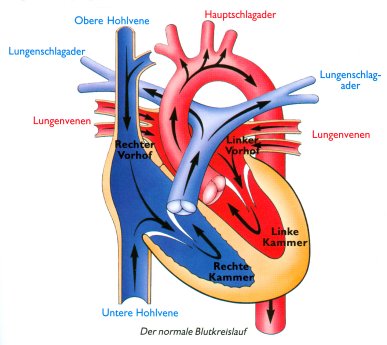
Brief overview of normal heart anatomy
In the healthy circulation, blood flows through the heart in an organized one-way pattern: from the body into the right atrium, then to the right ventricle, on to the lungs, back to the left atrium, through the mitral valve into the left ventricle, and from there through the aorta to the body.
When the mitral valve no longer closes normally, this organized flow is disrupted on the left side of the heart.
Which dogs are most commonly affected?
Older small-breed dogs are affected most commonly. Predisposed breeds include:
Commonly affected breeds:
Toy Poodle, Chihuahua, Miniature Schnauzer, Yorkshire Terrier, Dachshund, Cavalier King Charles Spaniel
Larger dogs are affected much less often, but they can also develop degenerative mitral valve disease.
What causes mitral valve disease?
The exact cause is not completely understood. The disease is believed to be related to degenerative changes in the connective tissue of the valve, with a strong genetic component.
Older theories suggesting that dental disease or bacterial spread causes the typical degenerative mitral valve changes are no longer considered the explanation for MMVD.
What is clear is that certain breeds are strongly predisposed and that the disease often runs in families.
What signs can occur?
In the early stage, the only finding may be a heart murmur. Clinical signs usually develop later.
- heart murmur
- coughing
- increased resting respiratory rate
- difficulty breathing
- reduced activity and exercise intolerance
- decreased appetite
- occasionally syncope or collapse
Not every dog with a murmur has immediate clinical signs. Many dogs remain stable for a long time before relevant heart enlargement or pulmonary edema develops.
How does the disease affect the heart?
Because the valve leaks, part of the blood moves backward into the left atrium during each heartbeat. At first, the body compensates for this through several mechanisms.
Over time, the chronic volume overload causes enlargement of the left atrium and later also the left ventricle.
As the disease progresses, cardiac function may deteriorate and fluid can build up in the lungs. This is called pulmonary edema.
Which additional complications can occur?
- cardiac arrhythmias
- pulmonary hypertension with possible strain on the right heart
- left atrial tear with bleeding into the pericardial sac, rare but life-threatening
- rupture of chordae tendineae, which can lead to sudden deterioration and acute pulmonary edema
These complications occur mainly in advanced cases.
How is the disease diagnosed?
Diagnosis is based on several examinations:
- physical examination and auscultation
- thoracic radiographs
- ECG
- echocardiography as the most important test
- blood pressure measurement in selected cases
The veterinary examination
Often the first finding is a heart murmur over the mitral valve area. The physical exam is the first step, but by itself it cannot reliably determine the stage of disease.
Radiographs
Radiographs are important to determine whether there is already heart enlargement or even pulmonary edema.
They are especially essential when evaluating possible congestive heart failure and fluid on the lungs.
ECG
ECG is mainly used to identify arrhythmias. Not every dog with MMVD needs an ECG at every visit, but it becomes important when rhythm abnormalities are suspected.
Echocardiography – the most important examination
Echocardiography shows valve changes, heart size and the severity of mitral regurgitation.
On ultrasound, the mitral valve can be evaluated directly. Typical findings include thickened, irregular valve leaflets and sometimes valve prolapse.
It also allows us to assess:
- size of the left atrium and left ventricle
- pumping function of the heart
- severity of mitral regurgitation with color Doppler
- possible signs of increased lung pressure
Depending on the case, advanced methods such as tissue Doppler, strain measurements or 3D echocardiography may also be used.
What is the prognosis?
Many dogs with mitral valve disease live stable lives for years. Prognosis depends strongly on whether there is already heart enlargement, clinical signs or pulmonary edema.
Once signs of decompensation appear, long-term prognosis becomes more guarded. However, early diagnosis, regular monitoring and individualized treatment can greatly improve quality of life.
What treatment options are available?
Treatment depends on disease stage, heart enlargement, clinical signs and associated problems.
Today, dogs with mitral valve disease are not treated only once pulmonary edema develops. Once there is relevant heart enlargement, treatment may already be recommended to slow progression of the disease.
Medication in the earlier stage
Once relevant heart enlargement is present, pimobendan is often prescribed. This can delay progression of the disease.
Medication when pulmonary edema or heart failure is present
- diuretics such as furosemide or torsemide to remove excess fluid and treat pulmonary edema
- pimobendan to support cardiac function
- ACE inhibitors in selected individual cases
- spironolactone or additional medications in more advanced disease
- antiarrhythmics when rhythm disturbances are present
At our clinic, treatment is always tailored to the individual patient. Regular follow-up is essential so medication can be adjusted in time.
Is there a surgical or interventional treatment?
Selected dogs with severe mitral regurgitation may be candidates for modern mitral valve repair procedures.
Open-heart mitral valve surgery is available only at very few centers. A promising minimally invasive alternative is TEER (transcatheter edge-to-edge repair) using the V-Clamp system.
This procedure is offered at the Small Animal Clinic of LMU Munich for selected dogs.
Learn more here: Mitral Valve Repair (TEER / V-Clamp)
Which dogs may be candidates for TEER?
Not every dog with mitral regurgitation is suitable for interventional repair. Important factors include:
- body weight
- disease stage
- functional and anatomic suitability of the mitral valve
- overall health status
Final assessment is always based on cardiologic examination and imaging.
What can owners do at home?
One of the earliest warning signs of developing pulmonary edema is an increased resting respiratory rate. Owners of dogs with mitral regurgitation should therefore monitor the breathing rate regularly at rest, ideally while the dog is asleep.
Normal: usually under 30 breaths per minute at rest
Above 30/min: monitor more closely
Around 40/min or higher: pulmonary edema becomes more likely and veterinary evaluation is strongly recommended
If the resting respiratory rate increases consistently or your dog shows coughing, labored breathing, weakness or reduced activity, a veterinary recheck is recommended.