Mitral Valve Repair in Dogs using the V-Clamp TEER Procedure
Advanced Mitral Valve Repair at LMU Munich
The Department of Veterinary Cardiology at the Small Animal Clinic of Ludwig-Maximilians-Universität München offers advanced minimally invasive mitral valve repair in selected dogs with severe mitral regurgitation caused by myxomatous mitral valve disease (MMVD).
The procedure is performed on the beating heart using the V-Clamp transcatheter edge-to-edge repair (TEER) technique. It is designed to reduce mitral regurgitation without the need for conventional open-heart surgery or cardiopulmonary bypass.
This page provides information for both dog owners and referring veterinarians.
Downloads
Download the owner information brochure for TEER surgery Download the PDF for veterinary cardiologists: required echocardiographic views for V-Clamp referralsWhat is Myxomatous Mitral Valve Disease (MMVD)?
 Myxomatous mitral valve disease, also called chronic mitral valve disease or mitral valve degeneration,
is a progressive degenerative disorder of the mitral valve. It is the most common cardiac disease in small dog breeds,
including Cavalier King Charles Spaniels, Dachshunds, Yorkshire Terriers and many other small breeds.
Myxomatous mitral valve disease, also called chronic mitral valve disease or mitral valve degeneration,
is a progressive degenerative disorder of the mitral valve. It is the most common cardiac disease in small dog breeds,
including Cavalier King Charles Spaniels, Dachshunds, Yorkshire Terriers and many other small breeds.
Degenerative changes in the valve tissue lead to thickening, shortening and loss of elasticity. As a result, the valve no longer closes completely and blood leaks backward from the left ventricle into the left atrium.
Main consequences of MMVD:
Mitral regurgitation: backward blood flow through the diseased valve
Volume overload: progressive enlargement of the left atrium and left ventricle
Heart failure: advanced disease may lead to pulmonary edema and clinical congestive heart failure
What is the TEER Procedure?
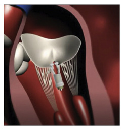
Transcatheter edge-to-edge repair (TEER) is a minimally invasive beating-heart procedure used to treat mitral regurgitation in selected dogs with degenerative mitral valve disease.
The V-Clamp procedure is the veterinary equivalent of the MitraClip concept used in human medicine. The device is placed under advanced imaging guidance in order to improve coaptation of the mitral valve leaflets and reduce valve leakage.
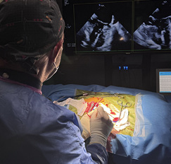
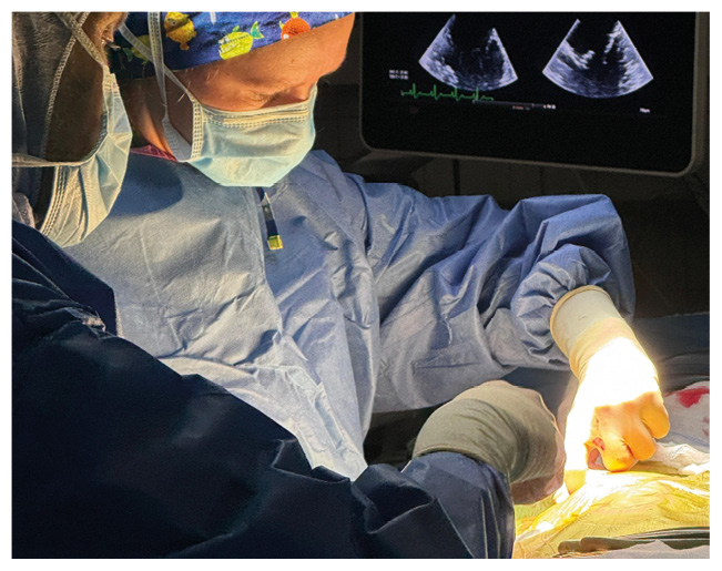
How is the V-Clamp Procedure Performed?
 The procedure is performed under general anaesthesia through a small thoracic incision.
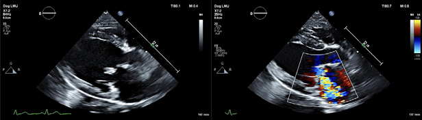
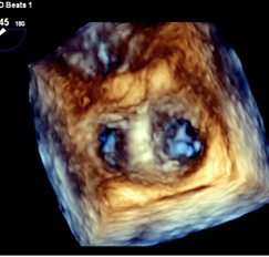
The V-Clamp device is guided into the beating heart using advanced imaging techniques,
especially 3D transoesophageal echocardiography.
The procedure is performed under general anaesthesia through a small thoracic incision.
The V-Clamp device is guided into the beating heart using advanced imaging techniques,
especially 3D transoesophageal echocardiography.
This approach is less invasive than conventional open-heart mitral valve repair and does not require a heart-lung machine.
Advantages of the TEER Procedure
- Minimally invasive approach through a small incision
- No cardiopulmonary bypass required
- Beating-heart catheter-guided procedure
- Short procedure time, typically around 40 to 60 minutes
- Usually short hospitalization and relatively fast recovery
- Tailored treatment because different device sizes are available
Clinical Outcomes and Long-Term Results
Long-term follow-up data for canine TEER have become increasingly encouraging and support the continued development of this procedure in carefully selected dogs.
In a large prospective study of dogs undergoing TEER, approximately three quarters of patients were still alive after one year, and around two thirds were still alive after two years. Freedom from cardiac-related death remained close to 80% at two years.
These results compare favorably with historical outcomes reported for advanced MMVD treated with medication alone, where survival after onset of congestive heart failure is often substantially shorter.
TEER is not suitable for every dog, and careful case selection remains essential. However, for selected patients with severe mitral regurgitation and suitable valve anatomy, this procedure may offer meaningful clinical benefit.
Is My Dog a Potential Candidate?
 Whether a dog is suitable for V-Clamp mitral valve repair depends on several factors and always requires
a detailed cardiological and imaging-based assessment.
Whether a dog is suitable for V-Clamp mitral valve repair depends on several factors and always requires
a detailed cardiological and imaging-based assessment.
A careful evaluation is essential to determine whether the expected benefits outweigh the procedural risks and whether the mitral valve anatomy is appropriate for repair.
Typical eligibility criteria include:
Clinical stage: late B2 or early C stage
Body weight: generally above 3 kg
Anatomy: suitable valve morphology for repair
General health: no major non-cardiac disease that would significantly increase anaesthetic or procedural risk
Initial Referral Assessment
 An initial assessment of eligibility is performed by the TEER / V-Clamp team at LMU Veterinary Cardiology
after echocardiographic examination.
An initial assessment of eligibility is performed by the TEER / V-Clamp team at LMU Veterinary Cardiology
after echocardiographic examination.
If your local cardiologist has already acquired the required echocardiographic views, we can review DICOM loops before your visit to Munich.
The final decision on procedural suitability is made under anaesthesia using transoesophageal echocardiography immediately before the intervention.
For referring veterinarians: please use the PDF with the required echocardiographic views and video loops: download referral echo views PDF.
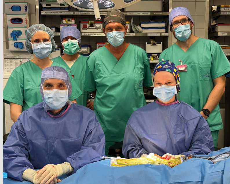
Why Refer to LMU Munich?
Mitral valve repair in dogs requires a highly specialized interdisciplinary team, advanced imaging expertise and substantial procedural experience. At LMU Munich, TEER is performed within a dedicated referral setting focused on interventional veterinary cardiology.
This includes collaboration between cardiology, surgery, anaesthesia and intensive care, as well as structured preoperative assessment and follow-up planning.
Risks of the Procedure
As with any cardiac intervention, complications can occur. The level of risk depends on the individual patient, anatomical suitability and the overall clinical situation.
Published clinical experience has shown good technical feasibility and encouraging outcomes, but TEER remains an advanced intervention and careful case selection is essential. Compared with conventional open-heart mitral valve surgery, the V-Clamp TEER procedure avoids cardiopulmonary bypass and is therefore generally considered less invasive.
Expected Benefits and Recovery
 The goal of TEER is to reduce the severity of mitral regurgitation and improve cardiac haemodynamics.
In many dogs, heart failure medication can be reduced after the procedure, although long-term cardiological follow-up
remains important and some dogs may require medication adjustments again over time.
The goal of TEER is to reduce the severity of mitral regurgitation and improve cardiac haemodynamics.
In many dogs, heart failure medication can be reduced after the procedure, although long-term cardiological follow-up
remains important and some dogs may require medication adjustments again over time.
Most patients can usually return home within 4 days after the intervention. Postoperative discomfort is typically limited, and mild pain medication may be required during the first few days.
Costs
The procedure currently costs approximately €12,000. If a second V-Clamp is required during the same operation, no additional device costs are charged.
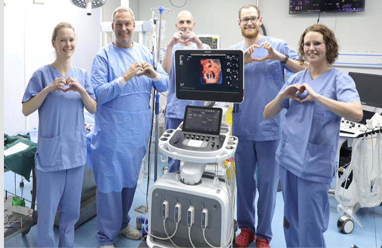
The LMU TEER Team
Our multidisciplinary TEER team at the Small Animal Clinic of LMU Munich, under the leadership of
Prof. Dr. Gerhard Wess, includes surgeons, cardiologists, a TEE imaging specialist as well as
dedicated support from anaesthesia and emergency / critical care.
This collaborative structure is essential for advanced mitral valve repair in veterinary patients.
Follow-Up Care
 Regular cardiological follow-up for at least one year after the procedure is strongly recommended in order
to monitor recovery and long-term heart function.
Regular cardiological follow-up for at least one year after the procedure is strongly recommended in order
to monitor recovery and long-term heart function.
Follow-up examinations may be carried out either at our clinic or by your local cardiologist, depending on the individual situation and location.
Referral and Case Evaluation
We offer case evaluation for referring veterinarians and international patients considering TEER.
For appointments, referral enquiries, DICOM submissions or case discussion, please contact the TEER team at LMU Munich.
Please send DICOM loops or a download link to: kleintier.kardiologie@lmu.de
General referral information can also be found here: Referral Information
Additional Brochure
The brochure below may also be useful for owners and referring veterinarians: