Pulmonic Stenosis in Dogs and Cats
What is pulmonic stenosis?
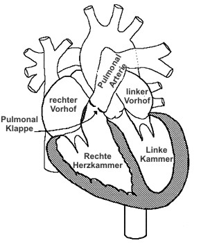
Pulmonic stenosis is a narrowing at the level of the pulmonary valve or the right ventricular outflow tract. The pulmonary valve is one of the four heart valves and regulates blood flow from the right ventricle into the pulmonary artery.
Because of this narrowing, the right side of the heart must pump against increased resistance. Depending on severity, this may lead to thickening of the right ventricular muscle, arrhythmias, and, in more advanced cases, clinical signs.
What causes pulmonic stenosis?
 The most common cause of pulmonic stenosis in dogs is a congenital malformation of the pulmonary valve.
The valve leaflets may be thickened, partially fused, and have reduced mobility.
The most common cause of pulmonic stenosis in dogs is a congenital malformation of the pulmonary valve.
The valve leaflets may be thickened, partially fused, and have reduced mobility.
As a result, blood flow from the right ventricle into the pulmonary artery is obstructed. The right heart must generate higher pressure to move blood across the narrowed area.
What types of pulmonic stenosis are there?
Narrowing can occur at different levels of the right ventricular outflow tract.
- Valvular pulmonic stenosis: narrowing directly at the pulmonary valve
- Subvalvular pulmonic stenosis: narrowing below the valve
- Supravalvular pulmonic stenosis: narrowing above the valve
In valvular pulmonic stenosis, different morphologic forms can occur. Clinically, it is important to distinguish between a more dome-shaped valve that still retains some mobility and a more severe valve dysplasia with thick, poorly mobile leaflets. This influences the likely success of balloon treatment.
Special form in some breeds
In Boxers and English Bulldogs, a special anatomic variant may occur in which an abnormal coronary artery encircles the pulmonary artery. This is particularly important because it can influence both the choice and safety of intervention.
Which breeds are more commonly affected?
In principle, pulmonic stenosis can occur in any dog or cat breed. In dogs, however, it is diagnosed more often in some breeds.
More commonly affected dog breeds:
French Bulldog, English Bulldog, Terrier breeds, Chihuahua, Miniature Schnauzer, Labrador Retriever, Mastiff, Chow Chow, Newfoundland, Basset Hound, Cocker Spaniel, Beagle, Samoyed
Which clinical signs may occur?
Many affected animals appear normal as puppies. In many cases, the disease is first suspected because a heart murmur is detected during a routine examination. Symptoms are more likely to occur in moderate to severe cases or when additional cardiac problems are present.
- exercise intolerance
- shortness of breath or exertional dyspnea
- arrhythmias
- syncope or collapse
- rarely sudden cardiac death
- in severe cases cyanosis
In advanced stages, right-sided heart failure with ascites, pleural effusion, or peripheral edema may develop.
How is pulmonic stenosis first detected?
The first clue is often a heart murmur heard during vaccination or a general physical examination. A typical murmur is a systolic murmur loudest over the left heart base.
Not every murmur in a puppy automatically means severe congenital disease. However, any persistent murmur should be evaluated further so that important congenital heart defects are not missed.
How is the diagnosis confirmed?
The most important examination for diagnosis and severity assessment is echocardiography.
ECG and thoracic radiographs may already provide supporting clues. However, the key examination is echocardiography with Doppler.
This allows the narrowing to be visualized, the effect on the right side of the heart to be assessed, and the pressure gradient across the stenosis to be measured.
2D echocardiography in pulmonic stenosis
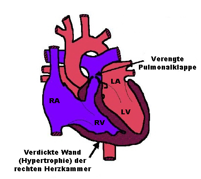
On two-dimensional ultrasound, the cardiologist can identify typical changes of the right heart. Because the right heart has to pump against increased resistance, a concentric hypertrophy of the right ventricle often develops.
Additional findings may include:
- narrowing at the valve level
- post-stenotic dilation of the pulmonary artery
- flattening of the interventricular septum
- reduced mobility of thickened valve leaflets
- dynamic obstruction of the right ventricular outflow tract
For prognosis and treatment planning, it is also important to assess whether there is marked valve dysplasia, annular hypoplasia, or other anatomic abnormalities.
Color Doppler and blood flow velocity
Color Doppler demonstrates the accelerated and turbulent blood flow across the stenosis. Typical mosaic color patterns indicate turbulent flow.
Based on the measured flow velocity, the pressure gradient across the stenosis can be calculated using the Bernoulli equation.
How is severity classified?
Severity is generally classified echocardiographically based on the pressure gradient.
Mild: 30–50 mmHg
Moderate: 50–80 mmHg
Severe: more than 80 mmHg
Dogs with mild pulmonic stenosis often have a normal life expectancy and normal exercise tolerance. In severe cases, the risk of clinical signs, syncope, and premature death is significantly higher.
How is pulmonic stenosis treated?
The earlier a clinically relevant pulmonic stenosis is recognized and treated when appropriate, the better the chance of limiting secondary changes in the heart.
In dogs with severe pulmonic stenosis, an intervention is usually recommended. In many cases, treatment is now performed by minimally invasive balloon valvuloplasty of the pulmonary valve.
In moderate cases, intervention may also be appropriate, especially when there is relevant tricuspid regurgitation or other evidence of clinically important right heart burden.
More detailed treatment information is available here: Balloon valvuloplasty for pulmonic stenosis
Prognosis
Prognosis depends strongly on severity, the anatomic type of stenosis, and the presence of additional cardiac changes. Mild disease often has a good long-term prognosis.
In moderate to severe cases, cardiology follow-up is important. In suitable patients, balloon valvuloplasty can significantly reduce pressure overload of the right heart.
What does balloon valvuloplasty cost?
The cost of balloon treatment for pulmonic stenosis at the Small Animal Clinic of LMU Munich is approximately 2,500 - 3000 euros and generally includes hospitalization, anesthesia, and the procedure itself.
Actual costs may vary depending on the individual patient, additional diagnostics, and material requirements.