Pulmonic Stenosis Balloon Valvuloplasty in Dogs and Cats
Minimally invasive treatment of a narrowed pulmonic valve

This page focuses on the treatment of pulmonic stenosis using balloon valvuloplasty. If you are looking for general information about the disease itself, symptoms, breed predispositions, and diagnosis, please also see our page on pulmonic stenosis in dogs and cats.
During balloon valvuloplasty, the narrowed pulmonic valve is dilated by a minimally invasive catheter-based procedure, allowing the right heart to pump blood more easily into the pulmonary artery.
When is balloon valvuloplasty recommended?
Treatment is mainly considered in patients with hemodynamically relevant pulmonic stenosis.
In patients with severe pulmonic stenosis, intervention is usually recommended. Balloon valvuloplasty may also be appropriate in selected moderate cases, especially when there are additional signs of right heart strain or significant tricuspid regurgitation.
Main goal: reduce pressure overload of the right heart
Typical procedure: balloon valvuloplasty via cardiac catheterization
Advantage: minimally invasive, without opening the chest
How is the procedure performed?
The intervention is performed under general anesthesia. Using fluoroscopic guidance, a catheter is advanced through a blood vessel in the neck or hind limb into the right heart.
A suitable balloon catheter is then positioned precisely across the stenotic valve and inflated under pressure. This can widen fused or obstructive portions of the pulmonic valve.
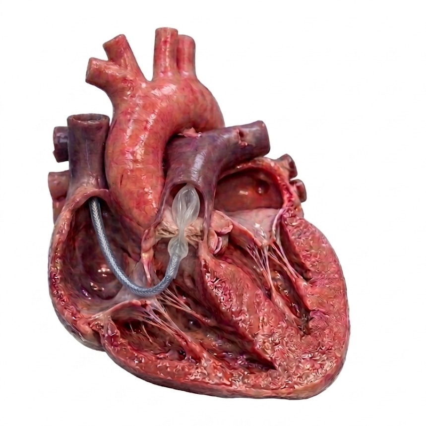
The catheter is introduced through the jugular vein and advanced into the right atrium, then into the right ventricle and across the stenotic valve.
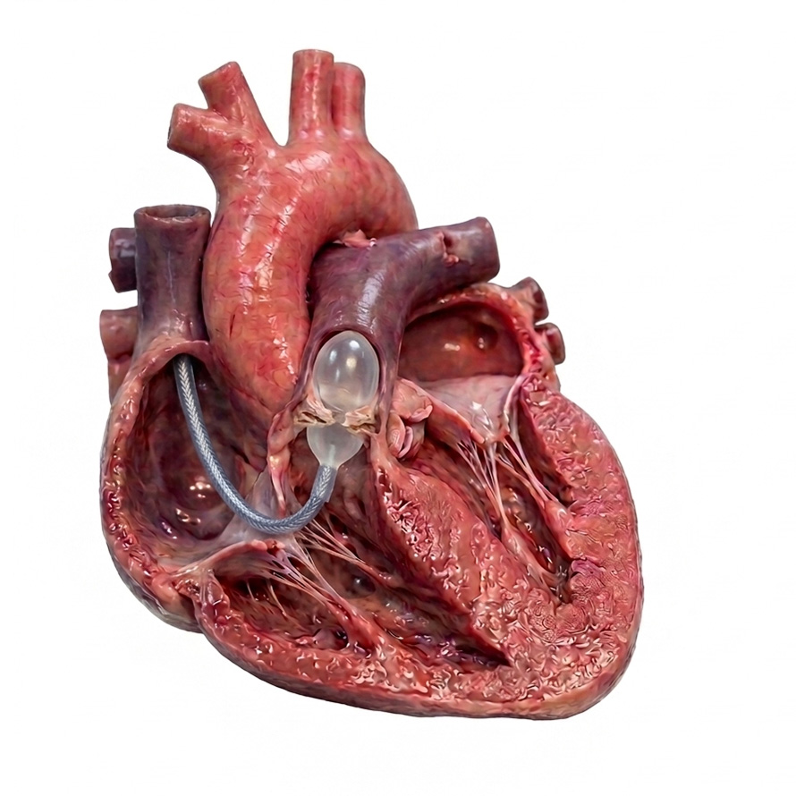
The balloon is positioned across the narrowed pulmonic valve and inflated.
What does balloon valvuloplasty achieve?
The aim is to reduce the pressure load on the right heart by improving opening of the narrowed valve.
After successful balloon valvuloplasty, the pulmonic valve may open more normally again, and in many patients the chronic pressure overload of the right ventricle is significantly reduced.
The best results are often seen in valves where fusion of the commissures is the main problem. In cases with marked valve dysplasia, thickened valve leaflets, and reduced mobility, the degree of improvement may be more limited.
Overall, many affected dogs show clear hemodynamic benefit after treatment.
Why experience matters
Balloon valvuloplasty for pulmonic stenosis is a specialized interventional procedure that requires experience in interventional cardiology, anesthesia, and perioperative monitoring.
At LMU Munich, these procedures are performed in a dedicated referral setting. This is important not only for the technical performance of the intervention, but also for patient selection and assessment of specific anatomical variants.
Transesophageal echocardiography during the procedure
When patient size allows, we also use transesophageal echocardiography during the intervention. This imaging technique allows us to evaluate cardiac anatomy in additional detail during planning and treatment.
This is especially helpful in breeds where special coronary artery anomalies may occur, for example in English or French Bulldogs.
Is the procedure risky?
As with any cardiac intervention, complications can occur, but balloon valvuloplasty is much less invasive than open-heart surgery.
Because the catheter is manipulated inside the heart, rhythm disturbances may occur, particularly in patients with severe right ventricular hypertrophy caused by advanced stenosis.
The procedure is therefore performed under close anesthetic supervision by an experienced team. Defibrillation paddles are also placed so that rapid treatment is possible if serious arrhythmias occur.
After the procedure
Following balloon valvuloplasty, patients are usually kept in hospital initially for monitoring. Depending on the individual case, heart rhythm, circulation, and echocardiographic findings are reassessed.
Further follow-up depends on the original severity of disease, valve anatomy, and the treatment result achieved.
Which patients benefit most?
The best candidates are usually dogs with valvular pulmonic stenosis in which fusion of the valve commissures plays a major role. In cases with marked valve dysplasia or additional anatomical abnormalities, the benefit may be less pronounced.
This is why a thorough cardiology examination before intervention is essential.
Estimated cost
The cost of balloon valvuloplasty for pulmonic stenosis is currently around EUR 2,500-3,000. This usually includes hospitalization, anesthesia, and the intervention itself.
Depending on the individual case, additional diagnostics or material requirements may lead to variation.
Further information
For referrals, questions, or appointments, please contact Veterinary Cardiology LMU Munich.