Cutting Balloon Intervention for Subaortic Stenosis in Dogs
Advanced interventional treatment for very severe subaortic stenosis
Cutting balloon intervention is a specialized catheter-based procedure used in selected dogs with very severe subaortic stenosis (SAS).
It is primarily considered in patients with extremely high pressure gradients and therefore an increased risk of syncope or sudden cardiac death.
This page focuses on the interventional treatment itself. General information about the disease is available on our subaortic stenosis page.
What is the goal of treatment?
The aim is to reduce obstruction in the left ventricular outflow tract and decrease pressure overload on the heart.
- reduce the pressure gradient
- decrease workload on the left ventricle
- lower the risk of syncope and sudden death in selected high-risk patients
When do we consider cutting balloon intervention?
Severe SAS: more than 80 mmHg
Particularly high risk: more than 130 mmHg
Dogs with very severe SAS, especially those with a pressure gradient of more than 130 mmHg, have a substantially increased risk of serious complications and sudden cardiac death.
In these patients, we recommend particularly close cardiology follow-up, appropriate medical treatment, and early discussion of whether interventional therapy may be beneficial.
From a medical standpoint, we now often use sotalol rather than atenolol in many of these patients. When it is clear that a dog is already in, or is likely to progress into, a very high gradient range, we also discuss the option of interventional treatment.
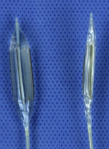
How does the cutting balloon procedure work?
The procedure is performed minimally invasively via cardiac catheterization.
- A catheter is advanced through a vessel to the heart.
- The narrowed region is first scored with a specialized cutting balloon.
- A high-pressure balloon is then used to dilate the prepared stenosis.
This approach may allow the fibrotic obstruction to be treated more effectively than with conventional balloon dilation alone.
Potential advantages over conventional balloon dilation
- more targeted disruption of the fibrotic narrowing
- better preparation for subsequent high-pressure balloon dilation
- potentially greater hemodynamic improvement in selected cases
Imaging during the procedure
The intervention is performed under fluoroscopy and also under advanced echocardiographic guidance.
We also use transesophageal echocardiography during the procedure. This allows the obstruction to be visualized and measured very precisely in both 2D and 3D.
Clinical experience at LMU Munich
Cutting balloon treatment has been performed at the Small Animal Clinic of LMU Munich for several years.
Although published long-term data remain limited, we now have meaningful practical clinical experience with this technique.
It may be especially useful in:
- dogs with very severe subaortic stenosis
- progressive disease courses
- patients with syncope or a high risk of sudden death
Is the procedure risky?
Like any interventional cardiac procedure, this treatment is not without risk.
Possible risks include:
- cardiac arrhythmias
- complications related to catheter manipulation
- anesthetic risk
For this reason, the procedure is performed under close anesthetic supervision, continuous ECG monitoring, and advanced imaging guidance.
Why should this be done at a specialized center?
Interventional treatment of subaortic stenosis requires extensive experience in interventional cardiology, anesthesia, and advanced imaging.
At LMU Munich, we perform complex catheter-based cardiac procedures on a regular basis, including cutting balloon interventions in dogs with very severe SAS.